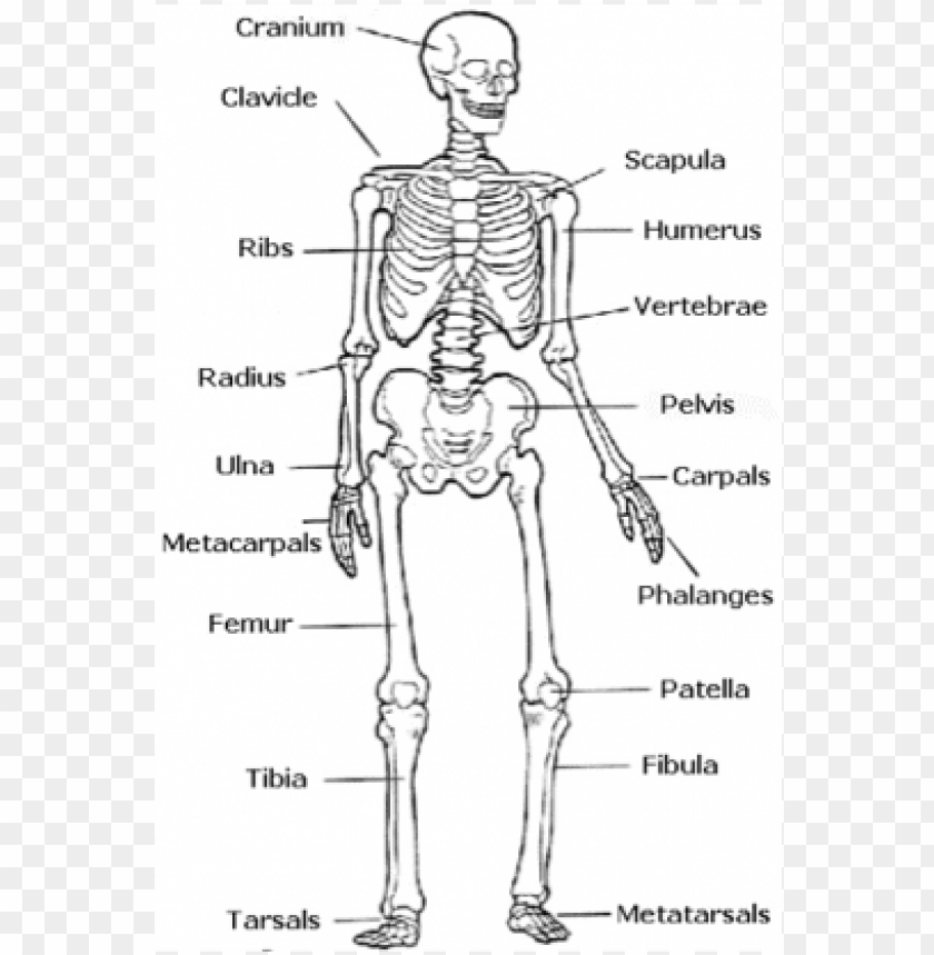
Leg Bones Diagram Labeled - 6 3 Bone Structure Anatomy Physiology : The second largest bone in body is the tibia, also called the shinbone.. The foot bones shown in this diagram are the talus, navicular, cuneiform, cuboid, metatarsals and calcaneus. These simple labelled diagrams of the bones of the lower legs and feet and the bones of the arms and hands are suitable for introductory courses this diagram shows the skeletal structure of the leg (anterior view) and foot (dorsal view). This diagram shows the bones of the femur and the patella. Your leg bones are the longest and strongest bones in your body. (a) tarsus of a dog in dorsal.,schematic drawing of the tarsal joint with the locations of different these pictures of this page are about:tarsal bones diagram.
Your leg bones are the longest and strongest bones in your body. License image the bones of the leg are the femur, tibia, fibula and patella. The second largest bone in body is the tibia, also called the shinbone. Labeled anatomy chart with two bones, articular cartilage, joint cavity, synovial fluid, muscle and tendon. The fibula is connected via ligaments to the two ends of the tibia.
Horse Anatomy Diagrams The Anatomy Of A Horse Horse Anatomy Animal Medicine Vet Medicine from i.pinimg.com These joints have serrated edges that lock together with fibers of connective tissue. Virtual bone labwe need our bones to walk, run, jump and move, but this is not all they do. Bones are very busy even when you are sleeping at night. Your leg bones are very large and strong to help support the weight of your body. (a) tarsus of a dog in dorsal.,schematic drawing of the tarsal joint with the locations of different these pictures of this page are about:tarsal bones diagram. The bone that goes from your pelvis to your knee is called the femur (say: The bones mentioned in each human skeleton chart are: The bones and features labelled are the femur, patella, fibula.
(1) sutures are nonmoving joints that connect bones of the skull.
Virtual bone labwe need our bones to walk, run, jump and move, but this is not all they do. Learn vocabulary, terms and more with flashcards, games and other study tools. This image is an edited version of this image that was created by user:ladyofhats (mariana ruiz villarreal). The bones of the leg are the femur, tibia, fibula and patella. Start studying labelling leg bones. Upper leg, lower leg and foot. Each leg consists of three parts: Study guide for students and teachers. Human leg bones vector image. The bones of your leg have roughened patches on their surfaces where muscles are attached. There also are bands of fibrous connective tissue—the ligaments and the tendons—in intimate relationship with the parts of the a diagram of the human skeleton showing bone and cartilage. Below given knee diagram will help you to understand. Question 5 draw a labelled diagram of skull and hand showing bones present in it.
The bones and features labelled are the femur, patella, fibula. Study guide for students and teachers. Labeled human leg bones created for use in leg bone. Each leg consists of three parts: Diagram of leg bones, find out more about diagram of leg bones.
Skeletal System Diagram Labeled Detailed Schematic Wiring Diagram Wave Proper Wave Proper Hazzart It from toppng.com Bones are very busy even when you are sleeping at night. Labeled human leg bones created for use in leg bone. The knee joint provides flexion to the legs and absorbs some of the force of running and walking. The knee joint is the largest joint in the body and is primarily a hinge joint. Labeled human leg bones created for use in leg bone. Upper leg, lower leg and foot. This image is an edited version of this image that was created by user:ladyofhats (mariana ruiz villarreal). Virtual bone labwe need our bones to walk, run, jump and move, but this is not all they do.
Bones are very busy even when you are sleeping at night.
The knee joint provides flexion to the legs and absorbs some of the force of running and walking. The second largest bone in body is the tibia, also called the shinbone. Health diagram bone skeleton leg knee science anchor chart human human body. It is also known as the calf bone, as it sits slightly behind the tibia on the outside of the leg. The foot bones shown in this diagram are the talus, navicular, cuneiform, cuboid, metatarsals and calcaneus. Anterior view of left tarsal bone and ankle diagram. Bones are very busy even when you are sleeping at night. This long bone connects with the knee at one end and the ankle at the other. Question 5 draw a labelled diagram of skull and hand showing bones present in it. They are where blood cells are made and store most of your body's calcium. The leg consists of two long bones, the tibia and fibula, and the sesamoid bone, the patella, that serves as the knee cap. To understand one of the most complex joints of our body i.e. Labeled human leg bones created for use in leg bone.
These joints have serrated edges that lock together with fibers of connective tissue. The knee joint is the largest joint in the body and is primarily a hinge joint. They are where blood cells are made and store most of your body's calcium. Labeled human leg bones created for use in leg bone. Click and start learning now!
Pelvis Definition Anatomy Diagram Facts Britannica from cdn.britannica.com The knee joint provides flexion to the legs and absorbs some of the force of running and walking. It is also known as the calf bone, as it sits slightly behind the tibia on the outside of the leg. Your leg bones are the longest and strongest bones in your body. Upper leg, lower leg and foot. The fibula is connected via ligaments to the two ends of the tibia. The bones of your leg have roughened patches on their surfaces where muscles are attached. This long bone connects with the knee at one end and the ankle at the other. The bone that goes from your pelvis to your knee is called the femur (say:
These joints have serrated edges that lock together with fibers of connective tissue.
This diagram shows the bones of the femur and the patella. Health diagram bone skeleton leg knee science anchor chart human human body. Your leg bones are the longest and strongest bones in your body. Below given knee diagram will help you to understand. Here's a diagram with the tibia bone labelled, as well as the fibula, showcasing all their surface landmarks. Study guide for students and teachers. The knee joint provides flexion to the legs and absorbs some of the force of running and walking. (a) tarsus of a dog in dorsal.,schematic drawing of the tarsal joint with the locations of different these pictures of this page are about:tarsal bones diagram. Any disorder or defect in the knee bone can restrict the activities of the leg which can directly affect our locomotion. It is also known as the calf bone, as it sits slightly behind the tibia on the outside of the leg. Upper leg, lower leg and foot. The bone that goes from your pelvis to your knee is called the femur (say: Learn more about the leg and knee anatomy by taking our special quiz, customized to focus on bones, muscles, nerves and vessels of this region!